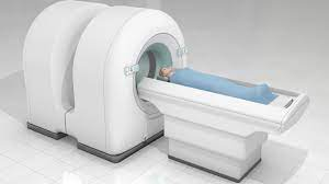
Is A Pet Scan Nuclear Medicine
Introduction
PET stands for positron emission tomography. This is a method of imaging the inside of the body using gamma rays that are produced by a radioactive sugar (or tracer) that becomes concentrated in cancerous cells. PET scans are often used to image tumors and other areas of concern, such as brain disorders like Alzheimer’s disease.
Section: Nuclear medicine uses very small amounts of radioactive tracers to image the body and treat disease. These tracers emit gamma rays that are detected by a specialized camera to create images of organs and tissues inside your body. PET scans can have lower image quality than an MRI or CT scan.
Section: A contrast agent is often used during a CT scan. Contrast agents injected into the body highlight abnormal tissue and make a clearer picture for the radiologist.
Section: A PET scan doesn’t usually require contrast because the radiation used for the scan is produced by a radioactive sugar that’s absorbed by all cells in the body, but most of it accumulates in cancerous cells.
Section: The use of radioactive materials in a PET scan may be a concern for some people, but most experts believe any risk from the radiation is outweighed by the benefits of early cancer detection.”
Takeaway: According to radiologyinfo.org, “the risks from having one or more PET scans are very low, since only very small amounts of radioactive materials are used..”
PET stands for positron emission tomography.
PET stands for positron emission tomography. A positron is the antimatter counterpart of the electron. To understand what a positron is, it’s helpful to know that all matter has both positive and negative charges. Electrons are negatively charged, while protons are positively charged. An electron can be thought of as a ball rolling around in orbit around a proton; however, if you replaced one of those protons with an anti-proton (a particle with similar mass but opposite charge), you’d end up with something called antimatter.
Positrons behave like regular electrons but have one important difference: they’re positively charged instead of negatively charged! When two particles or objects collide in space and annihilate each other, they release energy in the form of light—this phenomenon is called annihilation radiation (or pair production). When there’s enough energy present during this process, it becomes visible light (photons). In PET imaging machines that use nuclear medicine techniques such as PET/CT scanners or SPECT scanners like those used at Memorial Sloan Kettering Cancer Center have detectors designed specifically for detecting these photons from annihilation radiation emitted by radioactive isotopes injected into the patient being examined during his/her examination session.”

Nuclear medicine uses very small amounts of radioactive tracers to image the body and treat disease.
Nuclear medicine uses very small amounts of radioactive tracers to image the body and treat disease. Nuclear medicine is a medical imaging technique that uses small amounts of radioactive material to diagnose and treat disease. The images are not pictures of the body, but rather pictures of the tracer.
The advantages of using radionuclides for diagnostic imaging include their high sensitivity, specificity and accuracy; their ability to penetrate tissues that are normally opaque on conventional radiographs; the fact that they can be selectively bound to target organs or tissues (as in positron emission tomography); or their capability for generating signals after internalization by tumor cells (as with 99mTc-labeled compounds).
These tracers emit gamma rays that are detected by a specialized camera to create images of organs and tissues inside your body.
You will be in a comfortable position during the test, usually lying on your back.
A physician or technician will inject a small amount of radioactive tracer into your vein through an IV (intravenous) line that is inserted into one of your veins. The tracer emits gamma rays that are detected by a specialized camera to create images of organs and tissues inside your body.
PET scans can have lower image quality than an MRI or CT scan.
PET scans can have lower image quality than an MRI or CT scan. PET scans are more expensive than MRI and CT scans. PET scans are more accurate than MRI and CT scans. PET scans are more effective than MRI and CT scans. PET scans are more sensitive than MRI and CT scans
A contrast agent is often used during a CT scan.
Contrast agents are a type of substance used to make certain structures in the body visible on an X-ray or CT scan. They can be made from iodine, barium, or gadolinium. Contrast agents are injected into the body before the scan. This allows doctors to see better what is going on inside your body by making certain areas darker or lighter than they would have appeared otherwise.
Contrast agents injected into the body highlight abnormal tissue and make a clearer picture for the radiologist.
Contrast agents are substances that may be used in a medical imaging procedure to help the radiologist visualize certain structures. In CT scans, contrast agents are often used to highlight the abnormal tissue and make a clearer picture for the radiologist. Contrast agents can also be harmful or even fatal if injected into your body, so it is important to discuss with your doctor any risks associated with this type of treatment prior to receiving it.
A PET scan doesn’t usually require contrast because the radiation used for the scan is produced by a radioactive sugar that’s absorbed by all cells in the body, but most of it accumulates in cancerous cells.
- PET stands for positron emission tomography, which is also known as nuclear medicine.
- Nuclear medicine uses very small amounts of radioactive tracers to image the body and treat disease. These tracers emit gamma rays that are detected by a specialized camera to create images of organs and tissues inside your body.
- The radiation from a PET scan is similar to the natural radiation we’re exposed to in everyday life, such as from sunlight and cosmic rays. A PET scan usually doesn’t require contrast because the radiation used for the scan is produced by a radioactive sugar that’s absorbed by all cells in the body, but most of it accumulates in cancerous cells.
The use of radioactive materials in a PET scan may be a concern for some people, but most experts believe any risk from the radiation is outweighed by the benefits of early cancer detection.
When a PET scan is performed, the patient will be given an injection of a radioactive tracer substance. The area being examined is then scanned by the scanner, which detects radioactivity emitted from the tracer. This provides doctors and radiologists with information about how well oxygen is being delivered to that tissue.
In addition to being used for cancer detection, PET scans are also used to diagnose other diseases such as Alzheimer’s disease, heart conditions and epilepsy.
Because PET scans require radiation exposure, some people may be hesitant about having this test done if they’re concerned about potential health risks associated with nuclear medicine. However, most experts believe any risk from the radiation is outweighed by the benefits of early cancer detection and monitoring treatment progress over time
According to radiologyinfo.org, “the risks from having one or more PET scans are very low, since only very small amounts of radioactive materials are used.”
According to radiologyinfo.org, “the risks from having one or more PET scans are very low, since only very small amounts of radioactive materials are used.” In fact, the radiation exposure from these scans is so low that it is unlikely to cause any harm even if you have multiple scans (although there is still a small chance).
Another important consideration is that the benefits of early cancer detection far outweigh any risks associated with PET scanning. And because these scans can show up signs of cancer long before they become symptomatic—sometimes years before symptoms present—they may be quite useful in detecting cancers like breast and prostate cancer at an earlier stage when they’re easier to treat successfully.
Conclusion
I hope that you now have a better understanding of what a PET scan is, how it works and what to expect when having one. If you have any questions that weren’t covered in this article, please feel free to reach out to me or another member of our team. We would be happy to answer your questions about PET scans or anything else related to nuclear medicine.